Organ-on-a-Chip platforms can mimic the functions of individual organoids or entire organ systems. This is done by placing and maintaining tissue biopsies, 3D tissue models or cells within a microfluidic flow cell. Media can be pumped as desired and allowed to perfuse the cells with nutrients and oxygen, and then exit the system, removing waste products.
Organ-on-a-Chip technology has the potential to transform drug development. It constitutes an alternative to often unreliable animal models. A patient’s own tissue and cells can even be used in an Organ-on-a-Chip system, enabling personalised drug development.
In collaboration with biomedical researchers at the University of Hull, a range of devices for maintenance of tissue biopsies from brain, liver, gut and skin as well as cancer tissue were developed. Furthermore, there are examples of devices for spheroid maintenance and devices enabling the study of barriers.
Tissue Biopsy Devices
A skin-on-chip device was designed to model human skin biology and wound healing. The chip takes full thickness human skin biopsies, that are perfused with media at the bottom and have air/gas access at the top. The chips are reusable and can be cleaned and autoclaved [Kamil Talar PhD thesis and microTAS 2019].
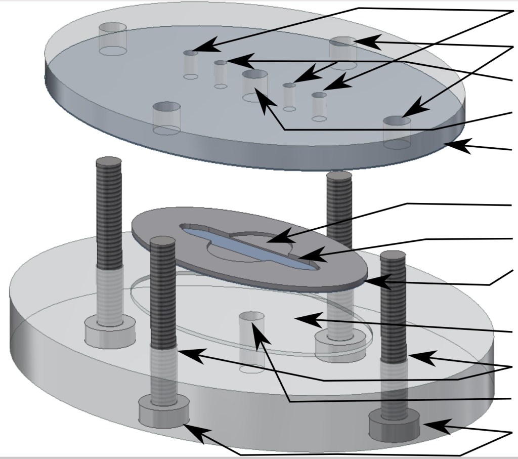
Our liver-on-a-chip device architecture was optimised using COMSOL simulations to enable the best possible mass transport of oxygen to a liver tissue slice to better simulate in-vivo oxygenation conditions. Two PDMS gas exchange membranes house the precision cut liver slice within a nylon mesh carrier. The chip provides fluidic control and continuous supply and removal of media, permitting effluent analysis. [MG Christensen, C Cawthorne, CE Dyer, J Greenman, N Pamme, Investigating oxygen transport efficiencies in precision-cut liver slice-based organ-on-a-chip devices, Microfl Nanofl, 2021, 25, 35. doi: 10.1007/s10404-021-02434-x]
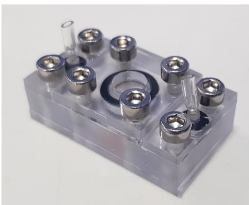
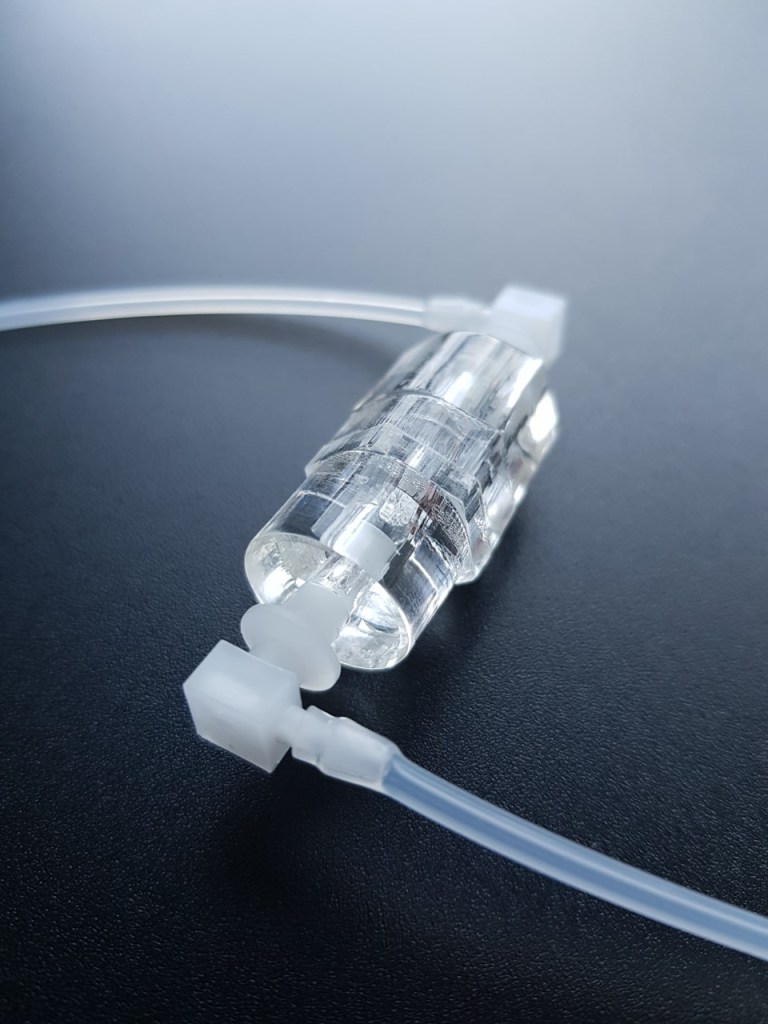
Example of a flow through device housing and maintaining tissue biopsies for perfusion. Such devices are employed in the groups of Dr Vicky Green and Prof. John Greenman at the Hull York Medical School. They are milled from polymer and interconnect to tubing through standard thread or push fittings.
Devices for Mimicking Barriers
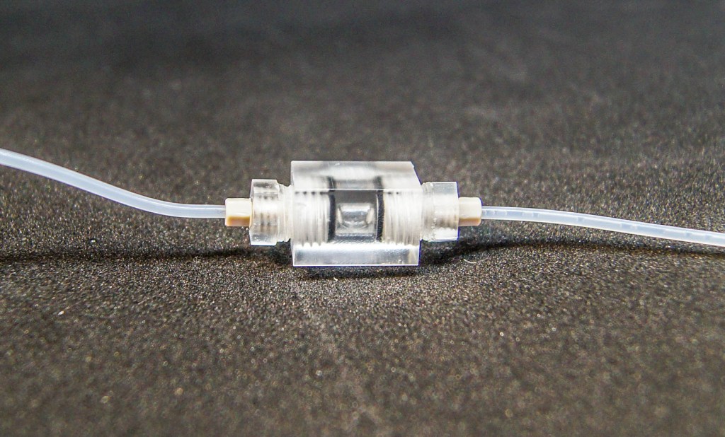
Dual flow devices feature an independent flow above and below a permeable membrane with cell cultures or a tissue biopsy held in a carrier. Such device designs can mimic barrier systems and thus act as gut-on-a-chip models or blood-brain-barrier models. The devices can also be daisy-chained for more complex multi-organ models. [PhD thesis Lydia Baldwin, microTAS 2019]
Spheroid Maintenance Devices
Spheroid are three-dimensional bundles of cells that simulate organ tissue more realistically than a two-dimensional layer of cells. Spheroids-on-chip can be used, e.g. to investigate cancer metastasis.
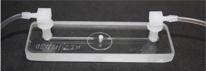
Here is an example of a glass microfluidic flow cell developed in collaboration with Dr Isabel Pires (University of Manchester) with chambers that allow placement, study and microscopic observation of spheroids. These have been applied to investigate the effects of hypoxia on cancer cells.
PhD thesis Emily Pyne and Thomas Collins, Emily Pyne, Martin Christensen, Alexander Iles, Nicole Pamme, Isabel M. Pires, Spheroid-on-chip microfluidic technology for the evaluation of the impact of continuous flow on metastatic potential in cancer models in vitro, Biomicrofluidics, 2021, 15, 044103. doi 10.1063/5.0061373.
In contrast, this flow-through spheroid device, developed in collaboration with Prof. Greenman’s team at the University of Hull, is fabricated from polymer and allows for study effluents. Fabrication cost is low such that the devices can be are employing this as a ‘single-use’ systems.
